
Getting a brain MRI can feel like stepping into a sci-fi movie. You lie still while a giant machine hums and clicks around your head. But for doctors, this image is the most powerful tool they have to see what’s happening inside your skull. It doesn’t use radiation, it shows incredible detail, and it helps diagnose everything from strokes to multiple sclerosis.
If you’ve ever looked at an MRI report or wondered why your doctor ordered one instead of a CT scan, you’re not alone. The images are complex, filled with black-and-white shades that mean different things depending on how they were taken. Understanding the basics can help you make sense of your results and ask better questions during your next appointment.
Why MRI Is the Gold Standard for Brain Imaging
Magnetic Resonance Imaging (MRI) has become the go-to method for investigating most central nervous system diseases. Unlike computed tomography (CT), which uses X-rays, MRI relies on strong magnetic fields and radio waves. This means no ionizing radiation, making it safer for repeated scans, especially in younger patients or those needing long-term monitoring for conditions like multiple sclerosis.
The real advantage lies in soft tissue contrast. An MRI can distinguish between gray matter and white matter with about 100 times more clarity than a CT scan. This is crucial when looking for subtle abnormalities. For example, in the posterior fossa-the back part of the brain where the cerebellum sits-CT scans often suffer from beam-hardening artifacts caused by bone. MRI avoids this issue entirely, providing a clear view of cranial nerves and small structures.
| Feature | MRI | CT Scan |
|---|---|---|
| Radiation Exposure | None | Yes (Ionizing) |
| Soft Tissue Detail | Excellent (100x better than CT) | Moderate |
| Scan Time | 30-45 minutes | 5 minutes or less |
| Best For | Chronic conditions, tumors, MS, stroke details | Acute trauma, bleeding, bone fractures |
| Cost (US Average) | $1,200 - $3,500 | $500 - $1,500 |
However, MRI isn’t perfect. It takes longer, which makes it less ideal for unstable trauma patients who need immediate answers. Also, people with certain metallic implants, like older pacemakers or cochlear implants, cannot undergo MRI due to safety risks. Despite these limitations, its diagnostic power keeps it at the top of the list for neurological investigations.
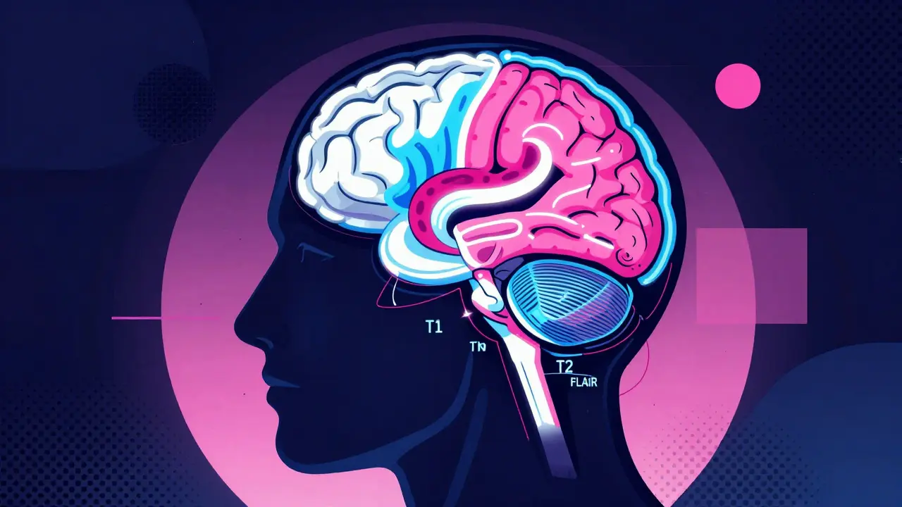
Decoding the Different MRI Sequences
An MRI isn’t just one picture; it’s a series of images called sequences. Each sequence highlights different properties of brain tissue by manipulating how water molecules respond to magnetic fields. Knowing which sequence shows what can help you understand why your radiologist picked specific images.
T1-weighted imaging provides excellent anatomical detail. In these images, fat appears bright white, while cerebrospinal fluid (CSF) looks dark. This makes it easy to see the structure of the brain, including the ventricles and cortical folds. If you’re looking at a T1 scan, think of it as a high-definition map of your brain’s geography.
T2-weighted imaging flips the script. Here, water-rich structures appear bright. This is useful because many pathological processes, such as edema (swelling) or inflammation, involve increased water content. However, since CSF is also water-based, it appears bright too, which can sometimes make lesions hard to distinguish from normal fluid spaces.
To solve this problem, radiologists use Fluid-Attenuated Inversion Recovery (FLAIR). FLAIR suppresses the signal from CSF, making it dark, while keeping pathological water signals bright. This is particularly valuable for detecting periventricular lesions in conditions like multiple sclerosis. If you see bright spots near the ventricles on a FLAIR image, that’s often where the pathology is hiding.
For acute emergencies, Diffusion-Weighted Imaging (DWI) is critical. DWI detects restricted water diffusion, which happens within minutes of a stroke. When combined with Apparent Diffusion Coefficient (ADC) maps, doctors can confirm an acute infarction if ADC values drop below 600 x 10^-6 mm^2/s. This speed allows for faster treatment decisions, potentially saving brain tissue.
Finally, Susceptibility-Weighted Imaging (SWI) or gradient echo sequences are sensitive to blood products. They can reveal microhemorrhages and calcifications that other sequences might miss. SWI is incredibly sensitive, capable of detecting hemosiderin concentrations as low as 0.5mg/dL, making it vital for assessing traumatic brain injury or vascular malformations.
Common Neurological Findings Explained
When you read an MRI report, you’ll likely encounter terms describing specific findings. Here’s what some of the most common ones mean:
- White Matter Hyperintensities: These are bright spots on T2 or FLAIR images. In older adults, they are often non-specific and related to aging or small vessel disease. However, in younger patients, they may indicate demyelinating diseases like multiple sclerosis. Location matters: lesions near the ventricles or in the corpus callosum are more suggestive of MS.
- Cerebral Atrophy: This refers to the loss of brain volume. On MRI, it appears as enlarged ventricles and widened sulci (the grooves on the brain surface). While some atrophy is normal with age, rapid progression can indicate neurodegenerative conditions like Alzheimer’s disease. Radiologists prefer using FLAIR images to assess atrophy accurately, avoiding overestimation that can occur on T2 images where bright CSF exaggerates ventricular size.
- Lacunar Infarcts: These are small strokes caused by blockages in tiny blood vessels. They appear as small, hyperintense (bright) foci on T2-weighted images, often in the basal ganglia or thalamus. Even if you don’t have symptoms, finding these can indicate a higher risk for future strokes or cognitive decline.
- Vestibular Schwannomas: Also known as acoustic neuromas, these are benign tumors on the hearing nerve. MRI is so sensitive it can detect tumors as small as 2mm in the internal auditory canal. If you have unilateral hearing loss or tinnitus, your doctor might look specifically at the cerebellopontine angle on your MRI.
- Flow Voids: These are dark areas within blood vessels where blood is moving too fast to generate a signal. Novice readers sometimes mistake them for pathology, but they are a normal finding. Recognizing flow voids prevents unnecessary worry about “black holes” in the brain.
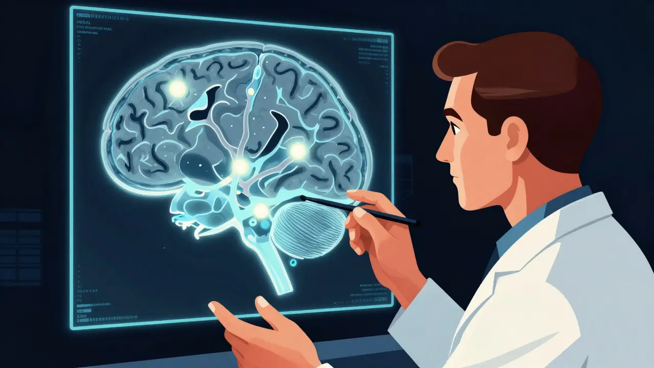
How Radiologists Interpret Your Scan
Radiologists follow a systematic approach to ensure nothing is missed. They typically start with midline structures and move outward. First, they check the ventricles for symmetry and size. Then, they examine subcortical structures like the basal ganglia and thalamus for signs of old infarcts or metabolic issues. Next, they review the brain lobes, cortex, meninges, and finally the skull.
Experience plays a huge role here. Studies show that radiology residents need 6-12 months of dedicated training to achieve 90% accuracy in identifying common pathologies. One key skill is distinguishing normal variations from disease. For instance, periventricular hyperintensities are present in 15% of people under 50 and up to 90% of those over 70. Without context, these could be mistaken for active disease.
Location clues are also vital. Temporal lobe involvement might suggest herpes encephalitis, while parietal-occipital predominance could point to posterior reversible encephalopathy syndrome (PRES). By correlating the image findings with your clinical history, radiologists provide a diagnosis that guides treatment.
When Is an MRI Necessary?
Not every headache needs an MRI. In fact, experts warn against overutilization. According to guidelines from the American College of Radiology, MRI is usually not appropriate for the initial evaluation of uncomplicated migraines without neurological deficits. Abnormal findings in such cases occur in only 1.3% of scans.
However, MRI is essential for specific scenarios. It is the gold standard for diagnosing temporal lobe epilepsy, characterizing brain tumors, evaluating infections like abscesses, and monitoring inflammatory conditions. If you experience sudden weakness, vision changes, confusion, or seizures, an MRI can provide the detailed information needed to plan your care.
Future Trends in Brain Imaging
Technology continues to evolve. Ultra-high field 7.0T MRI systems, cleared by the FDA in 2017, offer 0.5mm isotropic resolution, allowing visualization of individual cortical layers. While currently limited to academic centers, this technology promises deeper insights into neurodegenerative diseases.
Artificial intelligence is also changing the game. AI tools can reduce scan times by up to 50% while maintaining diagnostic accuracy. Additionally, advanced techniques like diffusion tensor imaging (DTI) for white matter tractography are becoming standard in multiple sclerosis protocols, helping doctors track disease progression more precisely.
Is a brain MRI safe?
Yes, brain MRI is generally safe because it does not use ionizing radiation. However, it involves strong magnetic fields, so it is not suitable for people with certain metallic implants like pacemakers, cochlear implants, or some aneurysm clips. Always inform your technician about any metal in your body before the scan.
What does a bright spot on an MRI mean?
A bright spot, or hyperintensity, depends on the sequence used. On T2 or FLAIR images, it often indicates increased water content, which can be due to inflammation, edema, scarring, or demyelination. In older adults, small bright spots are commonly associated with normal aging or small vessel disease. Context and location determine significance.
Can an MRI detect a stroke immediately?
Yes, specifically using Diffusion-Weighted Imaging (DWI). DWI can detect acute ischemic stroke within minutes of onset, far earlier than CT scans or conventional MRI sequences. This early detection is crucial for administering timely treatments like thrombolysis to restore blood flow.
Why did my doctor order an MRI instead of a CT scan?
Doctors choose MRI for its superior soft tissue contrast and lack of radiation. It is better for visualizing detailed brain structures, detecting small lesions, and evaluating conditions like multiple sclerosis or tumors. CT is preferred for quick assessments of acute trauma, bleeding, or bone fractures due to its speed and availability.
How long does a brain MRI take?
A standard clinical brain MRI typically takes 30 to 45 minutes. This time includes positioning, calibration, and acquiring various sequences. High-resolution studies or those requiring contrast injection may take slightly longer. Remaining still is important to prevent motion artifacts that could blur the images.